

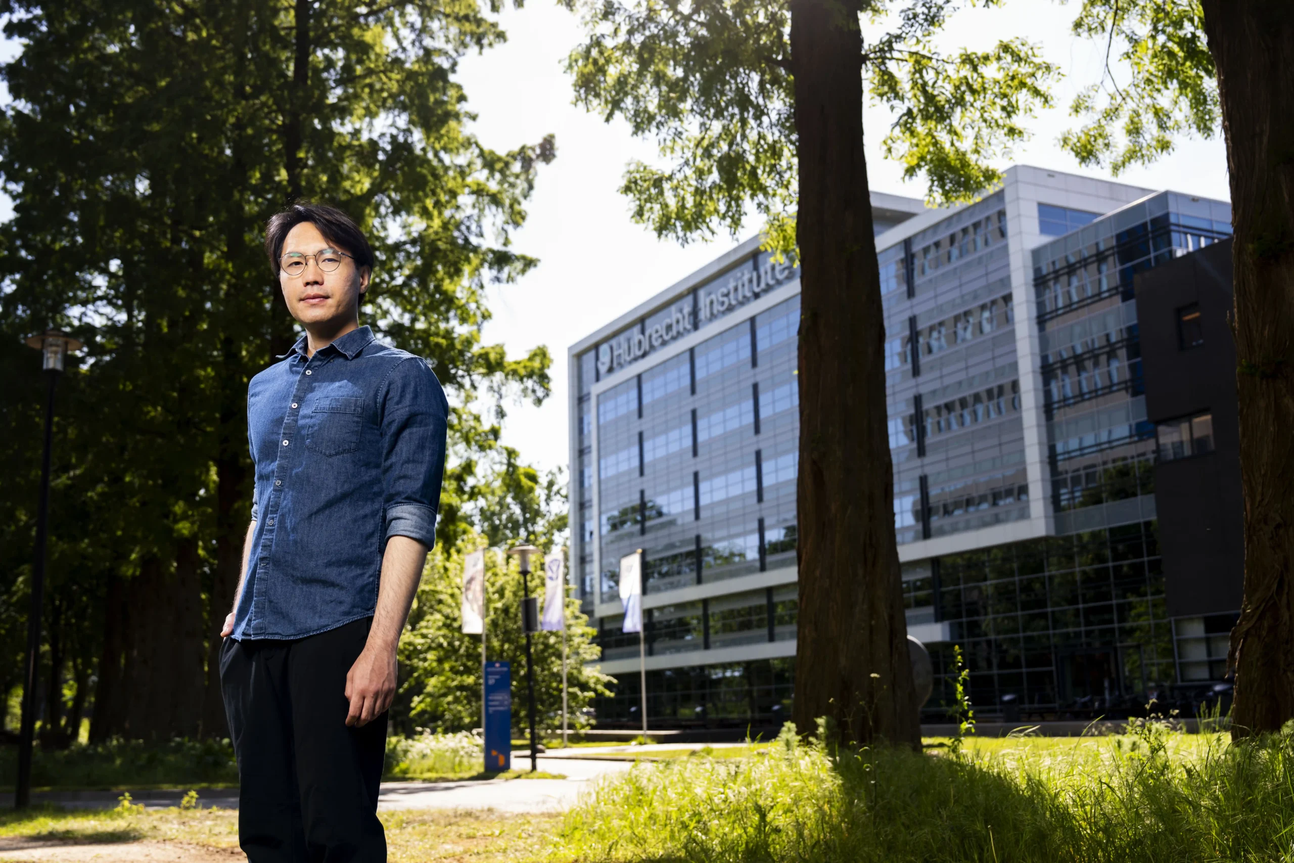
Miguel Leung (1996) — Hubrecht Institute
‘Unravelling the 3D structures of proteins tells us how the cell works’
The cause of many forms of infertility is still unknown. Something goes wrong with the sperm or egg cells, but exactly what is unclear. By closely studying the molecules in these cells, Miguel Leung is trying to unravel that mystery. As a structural biologist at the Hubrecht Institute, he uses advanced microscopy to map the three-dimensional structure of proteins. ‘Proteins are the molecules that do the real work,’ he explains. ‘This means that if we want to understand how cells function, we must first know what these proteins look like and how they interact. Only then can we determine what goes wrong in disease.’
Lees dit interview in het Nederlands (NewScientist)
Cilia and protein reserves
One of the key tasks of a sperm cell is to move towards the egg cell. It does this by moving its tail back and forth rhythmically. Within that tail is the molecular machinery which causes that movement. Together with his colleagues, Leung managed to map the 3D structure of this machinery, discovering that it consists of more than 150 different proteins. He also mapped the interactions between the proteins. ‘Many of these proteins were unknown to us because we simply did not have the methods to detect them,’ says Leung. ‘But now we have a complete overview and we can use it to potentially explain some forms of infertility that we do not understand yet.’
This type of swinging protrusion is not found solely in sperm cells. There are also various other places in your body that have cells with cilia that contain a similar molecular machinery for causing movement. There are cilia in your airways that propel mucus, for example, and your brain contains cilia that move cerebrospinal fluid. ‘We discovered that the propulsion system of sperm cells is by far the most complex in terms of structure,’ says Leung. ‘An interesting next step is to find out the reason why.’
When Leung took a closer look at the molecules in the egg, he managed to solve an important puzzle. ‘We already knew that mutations in certain egg cell proteins were known to cause infertility or disrupt embryonic development,’ he says. ‘But we did not know the exact function of these proteins.’ Earlier this year Leung and his team discovered that these proteins are part of an important structure: more than twelve proteins in the egg cell combine to form a huge storage complex. ‘Egg cells use this structure to create a protein reserve that is necessary for the early development of the embryo. In this case, something is probably going wrong with the storage of the reserve. Then we can take a look at where in the structure these mutations are located. This will help us gain a better understanding of how these problems are caused.’
Leung realised how little we actually know about systems that we thought we were already very familiar with. ‘People have been looking at sperm and egg cells for decades using other types of microscopic imaging,’ he says. ‘And many of the structures we study have already been studied. But by simply taking an even closer look, it is always possible to come across something unexpected. If you apply this to the endless diversity of life on our planet, I think we will never run out of research topics. As a young scientist, I see that as a wonderful realisation.’
Frozen proteins
Leung is able to examine huge quantities of interacting proteins in such detail thanks to cryo-electron microscopy and tomography. ‘The idea is to flash freeze a protein complex at incredible speed,’ says Leung. ‘This will basically make it stay trapped in the form it had inside the cell. We can then examine it with an electron microscope. We combine what we see through the microscope with artificial intelligence applications such as AlphaFold. This allows us to determine which proteins are in the complex.’
It used to be very difficult to create an image of a protein complex. Individual proteins had to be extracted from the cell and studied separately using complicated techniques. ‘But now we have the ability to visualise the proteins in close to their natural state, along with all their natural interaction partners,’ says Leung. ‘Whereas we used to have only a vague idea of what the proteins in the cell were doing, we can now see exactly which part of the protein interacts with which part of another protein. For example, in the case of disease, we can also see exactly whether incorrect protein interactions take place or if parts of proteins are missing.’
Seeing with your own eyes
The seed for a career in this field was planted early in Leung’s studies. ‘We were studying a famous publication by Watson and Crick during a course, in which they revealed the three-dimensional structure of DNA,’ he says. ‘I was struck by the elegance of this paper. It was a very short article, and just by knowing the structure of a molecule, you can learn so much about how it works. Watson and Crick concluded their article by stating that the structure they had found directly suggested a possible mechanism for copying the genetic material.’
Today, the structure of molecules is central to all of Leung’s research. ‘I often compare it to this: imagine you have read a lot about a certain place, and then you see it with your own eyes for the first time. Your idea of how that place is structured suddenly changes completely. In a sense, this also applies to structural biology. We currently still have a very abstract idea of how cells work. But if we suddenly knew what the proteins in those cells looked like, that would change the way we think about the problem. I hope that by making those structures visible, we can help all sorts of different researchers to think about their questions in new ways.’
Video
Young Scientist – Miguel Yeung